Before the white
chrysanthemum
the scissors
hesitate
a moment.
Yosa Buson
(1716 - 1784 / Japan)
Translated by Robert Hass
The seemingly strange name for this
plant derives from the Greek krus
anthemon which means gold
flower. Although chrysanthemums can be found today with
most of the colours of the spectrum, the first cultivated plants
mentioned by Confucius about 500 BC did have small yellow blooms.
Chrysanthemum cultivation is thought to have originated in China or
Japan. The Chinese considered it to be the ultimate flower, and
thus gave their throne the same name.
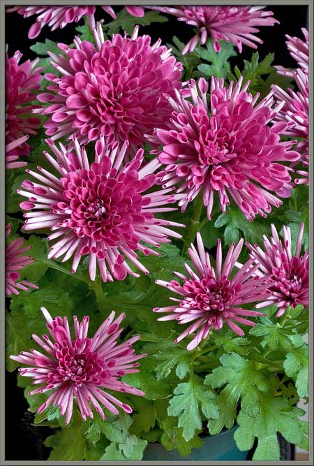
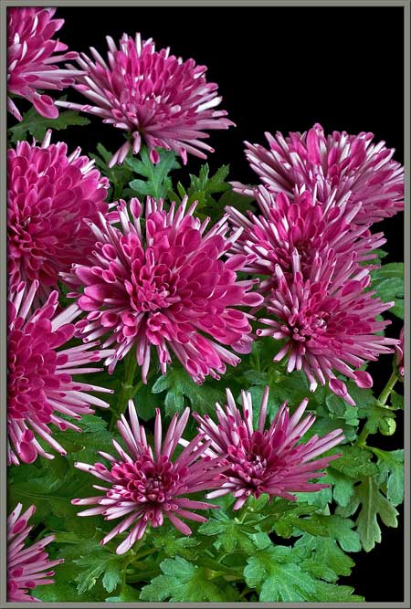
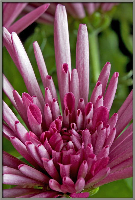
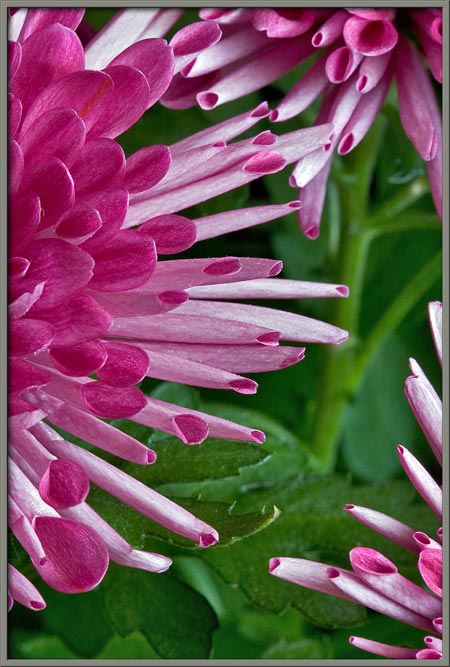
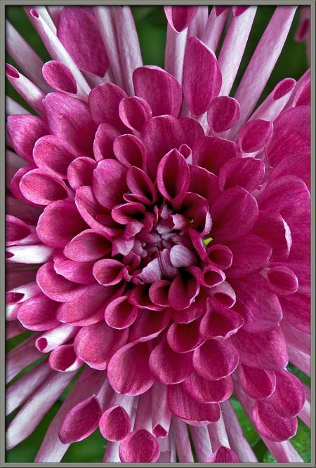
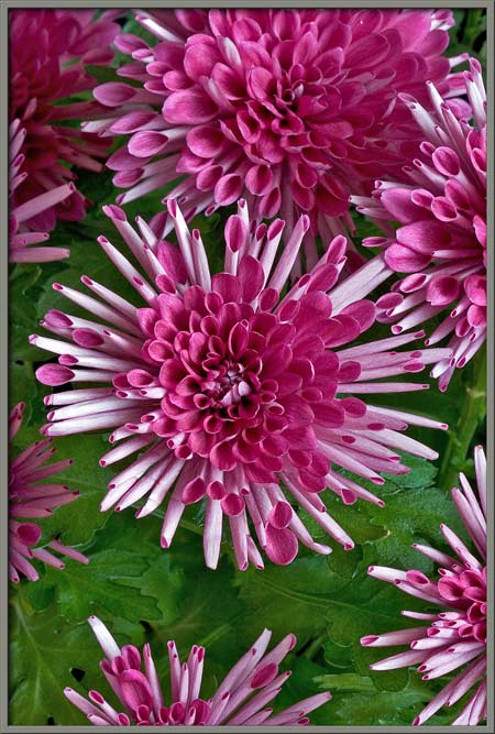
The flowers of a chrysanthemum are
typically clustered over the top of the plant where they may be so
abundant that they almost obscure the leaves beneath. Flowers of
different plants may possess a wide range of visual
characteristics. The petals may be daisy-like, narrow and lacy as
in the spider form, flat and spoon-shaped, feathery, or even
quill-shaped. The flower photographed for this article appears to
be a hybrid of quill and spoon forms.
I chose this particular plant
because of its striking colouration, with brilliant purple-red
spoons, and pinkish-white quills. Chrysanthemums belong to the
largest plant family on Earth the aster family (Asteraceae). Members of this
family usually possess both inner disk
flowers, and outer ray
flowers. Thus each of the blooms that can be seen below is
composed of many individual flowers (or florets).
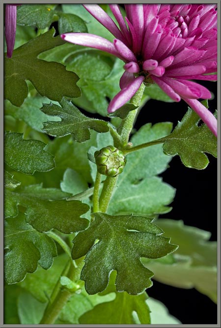
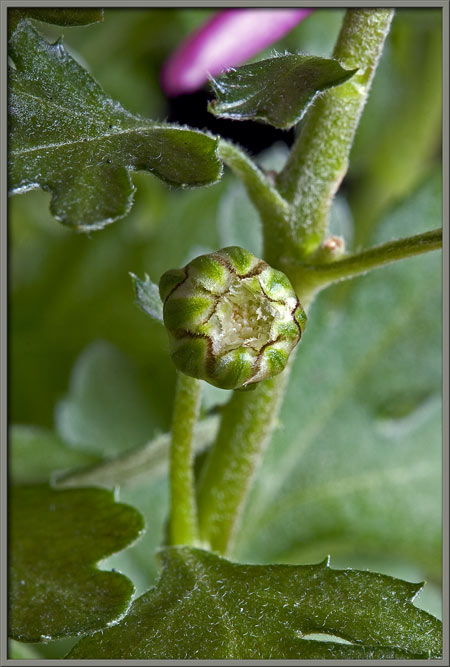
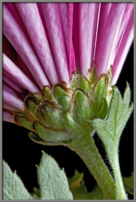
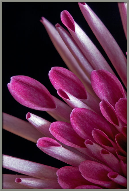
The three images that follow show a
chrysanthemum bud. Notice that the intense colour of the final
flower is nowhere to be seen in the bud form. The final image
shows clearly the ring of bracts
(modified leaves) that encloses the developing flowers. Notice
that the centre of each bract is green in colour, and that there is a
white band separating the centre from the dark brown edge.
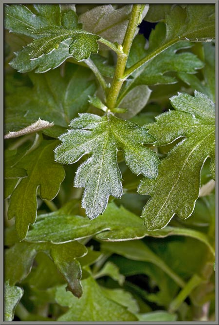
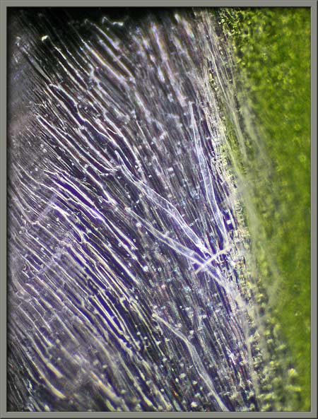
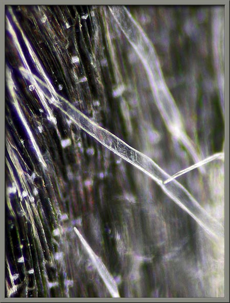
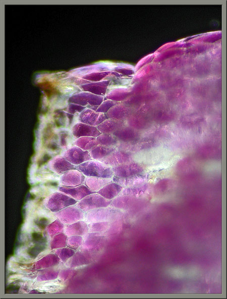
The green leaves are lobed and
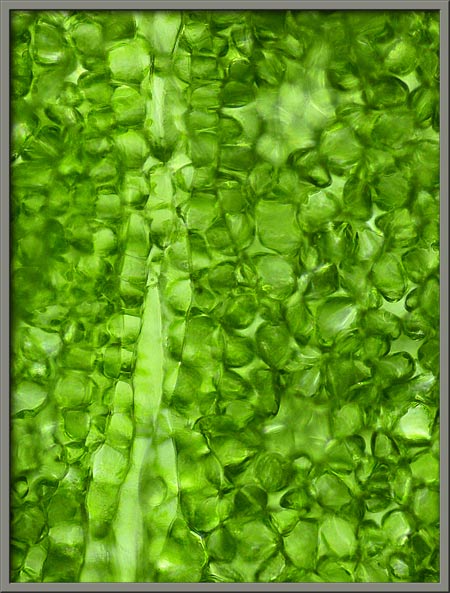
possess teeth around their margins. The photomicrograph at right
shows the cellular structure of the upper surface of a leaf.
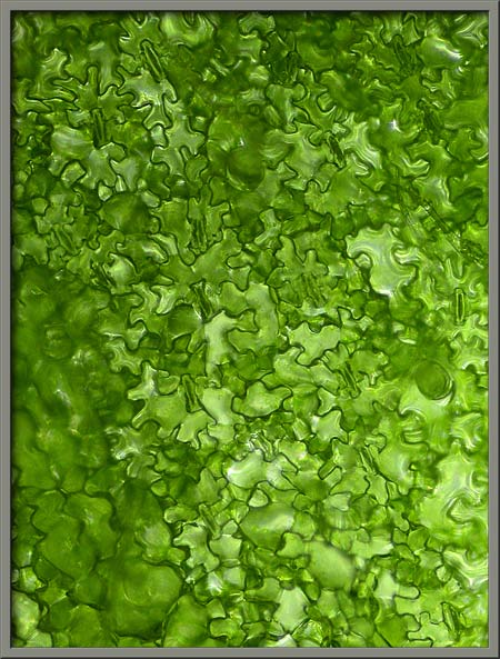
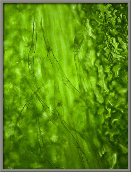
Each leafs lower surface is
composed of cells with a distinctive jigsaw appearance. Only the
lower surface possesses the soft hairs that can be seen in the
right-hand image.
At an early stage in the blooming
process, the outer quill-shaped disk flowers have yet to move into
their final position roughly perpendicular to the stem. Note the
multiple rings of bracts that enclose the bases of the flowers.
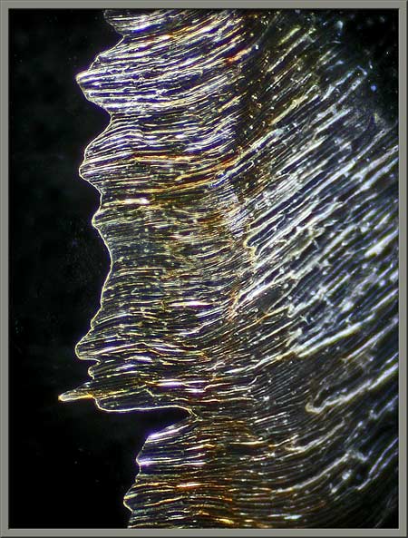
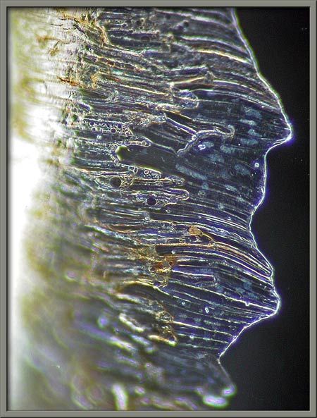
Under the microscope, the outer
edges of the bracts appear translucent, and have irregular shapes.
Some of the fine hairs that coat
the outer surface of the green middle section of a bract can be seen in
the photomicrographs below.
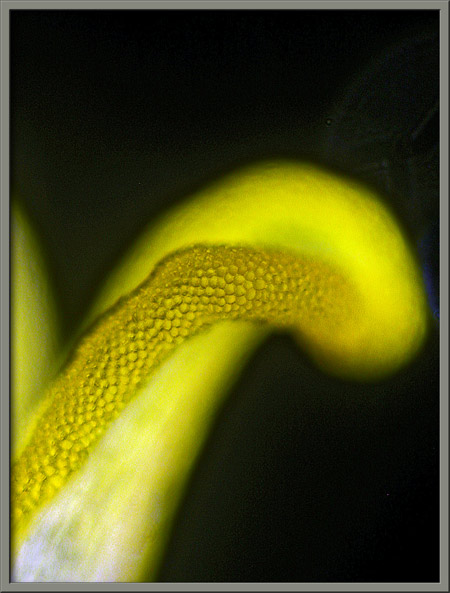
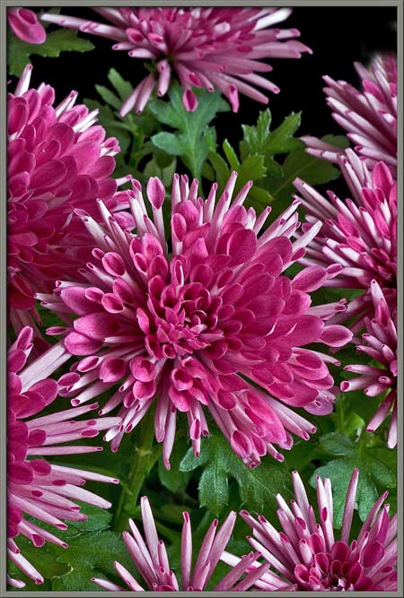
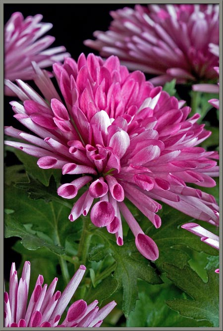
As mentioned earlier, the outer
disk flowers of this chrysanthemum have a long, tubular quill shape
with an oval opening at the tip. At the bottom end of each
tubular flower is the pistil,
(the female reproductive structure).
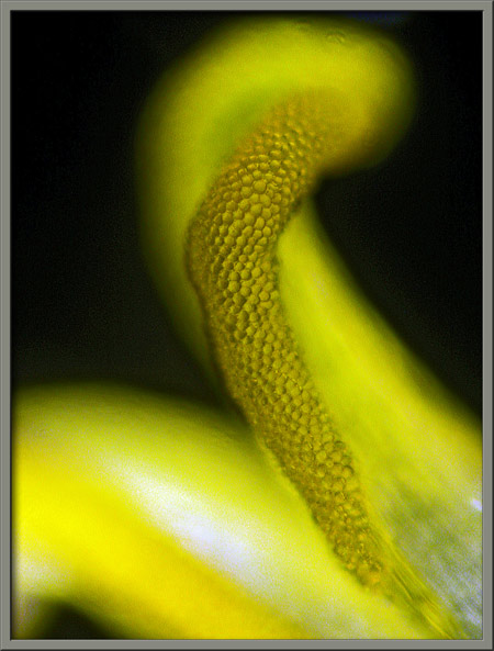
The three lobes of the stigma (the pollen accepting organ)
are initially parallel to one another (left image). Later, they
separate into the positions shown in the right-hand image. The style that supports the stigma can
be seen at the bottom of each image.
Higher magnification reveals that
the surface of each lobe is covered with dome-shaped protuberances.
Beneath the style is the
cylindrical ovary in which the flowers fruit, (or seed), forms. (The
style has been removed for this image.) The stalk that connects
the ovary to the base of the flower-head is visible in the image to the
right.
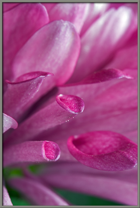
Between the outer ray flowers, and
the inner disk flowers, there is an intermediate ring of shorter quills
with spatulate (spoon-shaped) tips. These flowers contribute
greatly to the flower-heads striking appearance.
Notice how the edge of the quills
opening is rolled outwards.
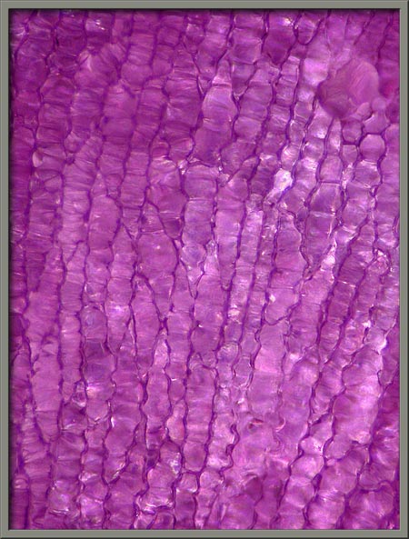
The two photomicrographs that
follow show the cellular structure of one of the spoon-shaped
petals. The image at left is true colour, while that on the
right was given a levels adjustment in Adobe Photoshop to enhance contrast, and
accentuate detail.
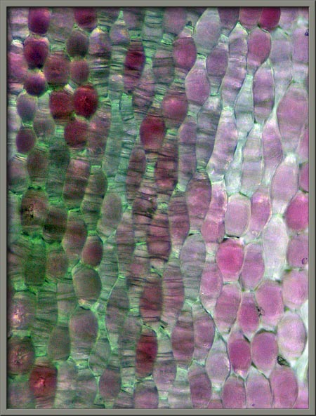
In the occasional petal, there is
some green colour along with the red. The higher magnification
image on the right shows the banded surface appearance of each cell.
Some of the cells at the topmost
edge of a quill can be seen below.
Each flower on the plant is subtly
different from the others. In the bloom below, there are
approximately equal numbers of quill and spoon flowers.
Other blooms have more spoon
flowers than quill flowers.
At the centre of the flower-head,
the tubular base of each spoon flower is so short that the flowers seem
to have simple oval petals.
Strangely, although I searched long
and hard for male stamens in the chrysanthemum flower-heads, I was
unable to find any. Perhaps an interested reader could give me a
hint as to where they might be found!
Although chrysanthemum blooms are
very diverse in structure, one can always identify the genus with the
aid of ones nose. Their unusual, and very distinctive scent
gives them away!
Photographic Equipment
The macro-photographs were taken
with an eight megapixel Canon 20D DSLR equipped with a Canon EF 100 mm
f 2.8 Macro lens which focuses to 1:1. A Canon 250D achromatic
close-up lens was used to obtain higher magnifications in several
images.
The photomicrographs were taken
with a Leitz SM-Pol microscope (using a dark ground condenser), and the
Coolpix 4500.
A Flower Garden of
Macroscopic Delights
A complete graphical index of all
of my flower articles can be found here.
The Colourful World of
Chemical Crystals
A complete graphical index of all
of my crystal articles can be found here.